
Jaw Cysts in Northwest Indiana: What You Need to Know
Jaw cysts are more common than you might think. These small, fluid-filled sacs can grow slowly in the bones of your mouth. Even though they are usually not dangerous, they can still cause problems with your teeth and jaw if left untreated. At Jay Platt DDS Oral Surgery & Dental Implant Center, we work to find and treat jaw cysts early to keep your mouth healthy.
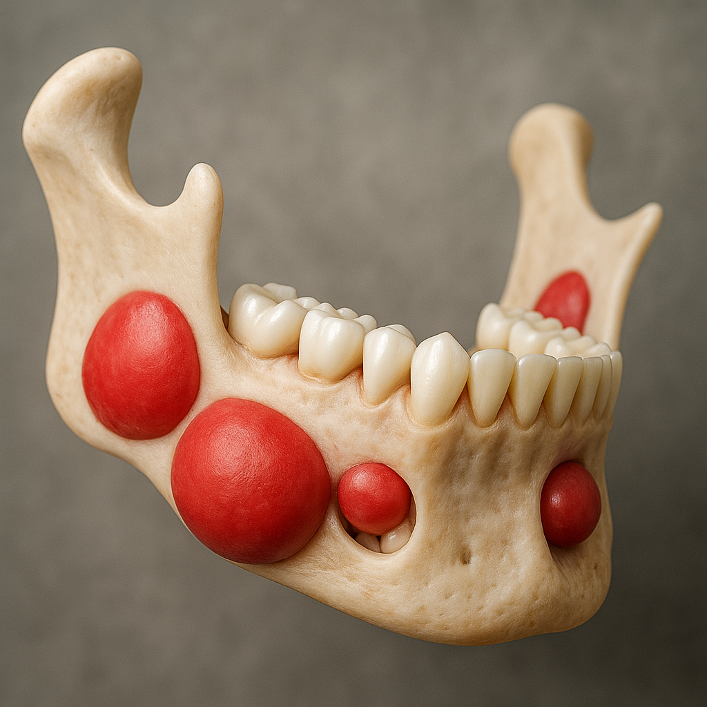
A jaw cyst is like a small bubble inside the bone of your upper or lower jaw. It’s filled with fluid or soft material and may not cause any pain at first. Many times, dentists find these cysts during regular X-rays. But if they grow too big, they can damage your jawbone, push teeth out of place, or even cause part of your jaw to break.1
Types of Jaw Cysts
One type is called a dentigerous cyst. It grows around a tooth that hasn’t come in yet, like a wisdom tooth. It’s the second most common kind of jaw cyst.2
A radicular cyst is the most common. It happens when a tooth has been infected for a long time and the tissue around the root gets inflamed.3
Another kind, called an odontogenic keratocyst (OKC), grows more quickly and often comes back after being removed. Doctors now call it a tumor because of how it acts. It’s sometimes linked to a genetic condition called Gorlin-Goltz syndrome.4
Other cysts include residual cysts, which stay behind after a tooth is taken out, and nasopalatine duct cysts, which grow behind the front teeth at the top of your mouth. There are also a few rare types that can show up in the jaw.
Causes and Risk Factors
These cysts can be caused by infections, problems during tooth development, or injury. Teeth that don’t grow in properly, predisposing health issues, not brushing and flossing well, or ignoring dental problems can also raise your risk.
Symptoms
At first, you might not feel anything at all. But as a cyst gets bigger, you may notice:
- Swelling or a lump in your jaw
- Pain or tenderness
- Teeth that feel loose
- Numbness or tingling in your face
- A delay in a tooth coming in (especially in kids)
Diagnosis
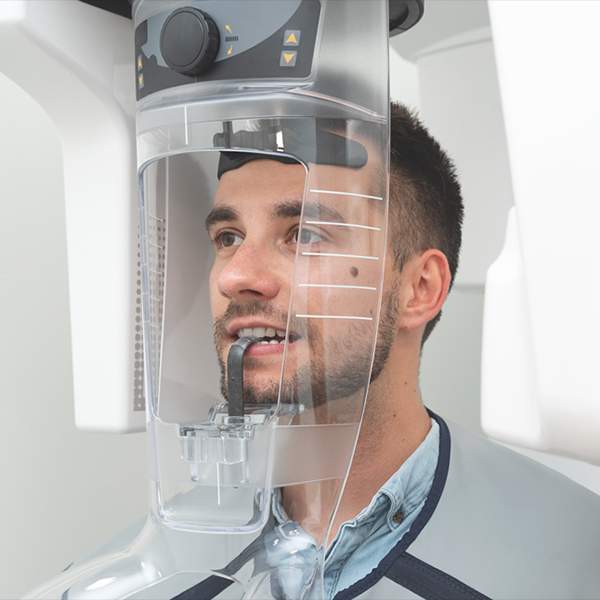
Dentists use special X-rays or 3D scans called cone beam CT to look for jaw cysts. If they find something unusual, they may take a small sample, called a biopsy, to figure out what it is.
Treatment
Doctors usually remove cysts with surgery. The two main ways to treat jaw cysts are:
- Enucleation – This means the whole cyst is taken out during surgery.5
- Marsupialization or Decompression – This is a gentler way to treat bigger cysts. The doctor makes a small opening in the cyst so it can shrink over time. Later, they may go back and remove the rest. This method works well for children and when the cyst is close to important areas.6,7
For larger or fast-growing cysts like OKCs, doctors may use both methods in steps to get better results and lower the chance of the cyst coming back.7,8
After surgery, follow-up appointments are important to make sure the cyst doesn’t return, especially for types like OKCs and glandular cysts.9
When to See an Oral Surgeon
If you notice any unusual swelling, pain, or changes in your teeth or jaw, it’s a good idea to see a specialist. If your dentist spots something odd on an X-ray, they may refer you to someone like Dr. Platt. Acting early can help prevent bigger problems later.
Read Next: Osteonecrosis of the Jaws: Should You be Worried?
Related Readings
What to Expect Before and After Oral Surgery with Dr. Jay Platt
Spotting the Silent Threat: Why Regular Oral Cancer Screenings Are a Lifesaver
Detecting and Preventing Oral Cancer: The Importance of Oral Surgeons
References
- Shear M, Speight P. Cysts of the Oral and Maxillofacial Regions. 4th ed. Oxford, UK: Wiley-Blackwell; 2007.
- El-Ghouzzi R, El-Hassani K. Dentigerous cyst: enucleation or marsupialization? A case report. Pan Afr Med J. 2021;40:149. doi:10.11604/pamj.2021.40.149.31513
- Nair PN. Non-microbial etiology: periapical cysts sustain post-treatment apical periodontitis. Endod Topics. 2003;6(1):96-113. doi:10.1034/j.1601-1546.2003.00042.x
- Madras J, Lapointe H. Keratocystic odontogenic tumour: reclassification of the odontogenic keratocyst from cyst to tumour. J Can Dent Assoc. 2008;74(2):165e. PMID: 18353202.
- Bodner L. Cystic lesions of the jaws in children. Int J Pediatr Otorhinolaryngol. 2002;62(1):25-29. doi:10.1016/S0165-5876(01)00625-4
- Zhao YF, Wei JX, Wang SP. Treatment of odontogenic keratocysts: a follow-up of 255 Chinese patients. Oral Surg Oral Med Oral Pathol Oral Radiol Endod. 2002;94(2):151-156. doi:10.1067/moe.2002.125400
- Pogrel MA. Decompression and marsupialization as a treatment for the odontogenic keratocyst. Oral Maxillofac Surg Clin North Am. 2003;15(3):415-427. doi:10.1016/S1042-3699(03)00043-4
- Tolstunov L, Treasure T, Oshima T. Surgical treatment algorithm for odontogenic keratocyst: combined treatment of decompression and enucleation with peripheral ostectomy. J Oral Maxillofac Surg. 2016;74(3):535-542. doi:10.1016/j.joms.2015.10.019
- Ide F, Mishima K, Saito I, Kusama K. Glandular odontogenic cyst: a comprehensive review. J Oral Maxillofac Surg. 2013;71(7):1304-1310. doi:10.1016/j.joms.2012.12.013
Comments are closed.